What you’ll learn
- Explain negative feedback through a named variable and corrective response.
- Connect insulin and glucagon to appropriate tissues and processes.
- Distinguish ultrafiltration, selective reabsorption and ADH-controlled water permeability.
Connections: AQA 3.6.4 · OCR A 5.1.1, 5.1.2, 5.1.4. Selected glucose and water-potential regulation and kidney mechanisms. Thermoregulation, detailed liver metabolism, pancreatic ion-channel secretion, glucagon second-messenger detail, renal treatments and complete endocrine signalling require further study.
In this chapter
Negative feedback
Homeostasis maintains internal conditions within ranges compatible with cell function. Receptors detect changes, a coordinating system receives the information, and effectors act on the condition being regulated. In a glucose-control question, name blood glucose as the variable and explain how the response changes it.
In negative feedback, the response opposes the departure that initiated it. A rise can activate a process that lowers the variable, while a fall can activate a process that raises it. The response can diminish as the departure is corrected. This is not the same as positive feedback, where a change promotes further change in the same direction, as during the rapid depolarising phase of an action potential.
A controlled condition can fluctuate; homeostasis does not mean an absolutely constant reading. Meal timing, activity and other signals also influence glucose regulation. A graph may show a rise after a meal followed by a delayed corrective response. That rise alone does not show that regulation has failed; follow what happens next.
Blood glucose regulation
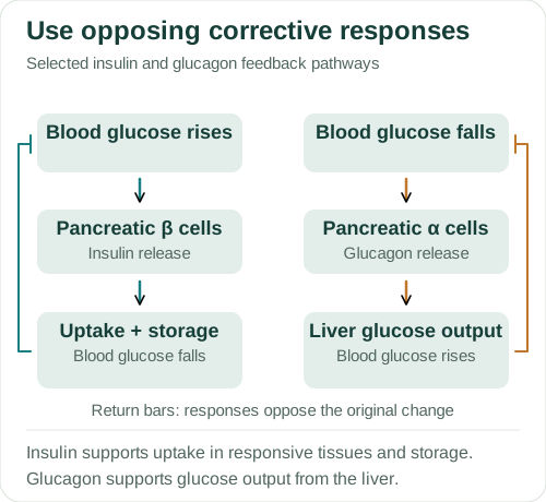
A rise in blood glucose promotes insulin secretion by pancreatic β cells. Insulin binds receptors on responsive tissues and changes their activity. In skeletal muscle and adipose tissue, it promotes insertion of glucose transporters into the cell-surface membrane, increasing uptake under the appropriate gradient. In liver and muscle it supports glycogen synthesis; other effects influence glucose use and storage.
Liver cells handle glucose differently from skeletal muscle cells. Insulin affects liver glucose handling through metabolic regulation, including storage and reduced glucose output. Glucose crossing through a facilitated-diffusion transporter is not itself an ATP-driven pump step, even though the signalling and metabolism have energetic requirements.
A fall in blood glucose promotes glucagon secretion by pancreatic α cells. Glucagon acts chiefly on the liver, stimulating glycogen breakdown and supporting gluconeogenesis. These processes increase hepatic glucose output. Muscle glycogen primarily fuels the muscle’s own metabolism. It does not directly supply free glucose to maintain blood glucose in the way liver glycogen does.
Type 1 and type 2 diabetes involve different failures of glucose regulation. Type 1 involves insufficient insulin production; in type 2, tissues respond less effectively to insulin, and insulin secretion can also become impaired.
About this diagram
Selected negative-feedback pathways, not all glucose-control mechanisms. Tissue responses differ: insulin-dependent transporter insertion concerns responsive tissues such as skeletal muscle and adipose tissue; liver glucose handling has additional mechanisms. No fixed glucose set point or clinical treatment is implied.
Ultrafiltration at the glomerulus
The liver converts nitrogen from amino-acid breakdown into urea, which is transported in blood to the kidneys. In a nephron, the glomerulus is a capillary network within Bowman’s capsule. Pressure drives filtration across a selective barrier into the capsule. Water and many small dissolved substances, including urea, glucose and ions, can enter the filtrate; blood cells and most large plasma proteins remain in the blood.
Ultrafiltration is not a sorting process that selects only wastes. Useful small molecules are filtered too. The barrier’s structure and molecular properties constrain passage, while the net filtration pressure also depends on pressures across the barrier. After filtration, tubular transport changes the composition of the fluid as it flows through the nephron.
Refer to the fluid in Bowman’s capsule and tubules as filtrate or tubular fluid. It does not already have the composition of final urine. Final excretion reflects filtration, reabsorption and secretion together. A substance can be filtered but largely recovered, so “it passes the filter” does not establish that it will be excreted in large amounts.
Selective reabsorption
In the proximal convoluted tubule, epithelial transport recovers glucose and other useful substances under ordinary physiological conditions. Sodium–potassium pumps on the blood-facing membrane help maintain a low intracellular sodium concentration. Sodium can enter from the lumen through coupled transporters, driving uptake of substances such as glucose. Glucose can then leave towards the surrounding fluid and blood through a suitable carrier.
Water follows favourable water-potential gradients by osmosis, while different ions and solutes have their own routes. Microvilli provide area for transport, and mitochondria supply ATP needed for processes including pumping. The extra membrane area accommodates transport proteins, while the ATP supply allows pumps to maintain the gradients used in reabsorption.
Transport capacities are finite. When filtered glucose delivery exceeds the relevant reabsorptive capacity, some remains in the tubular fluid and is excreted. To calculate recovery, compare the filtered and excreted amounts and check whether secretion also contributes.
The loop of Henle
The descending limb of the loop of Henle is relatively permeable to water. As tubular fluid descends into the increasingly concentrated medulla, water can leave by osmosis. The ascending limb has low water permeability; salt movement out of the limb, including active transport in its thick region, contributes to the medullary concentration gradient.
Opposite flows in the two limbs help maintain and multiply the gradient along the medulla. The resulting low medullary water potential allows water to leave the collecting duct when its walls are permeable. The whole concentrating mechanism also involves vascular exchange and urea recycling, which are not drawn in the collecting-duct figure.
An impermeable ascending limb cannot lose water by the same mechanism as a permeable descending limb. Nor does a water channel itself create the gradient. Keep permeability and driving force separate: net osmosis requires both a route and a water-potential difference.
ADH and water reabsorption
A fall in blood water potential is detected by osmoreceptors in the hypothalamus. ADH is synthesised in hypothalamic neurones and stored and released from the posterior pituitary. Increased ADH release can act on collecting-duct cells, making the lumen-facing membrane more permeable to water.
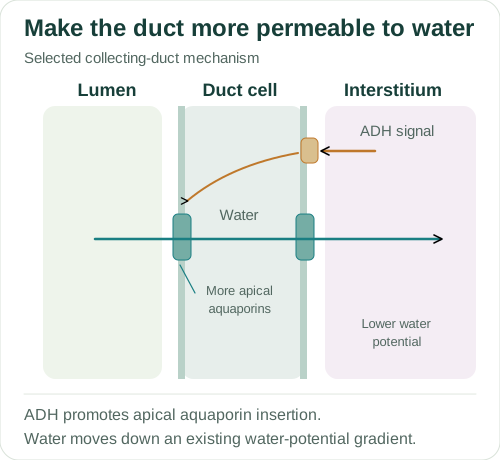
ADH binds receptors on the blood-facing side of a collecting-duct cell. Signalling promotes insertion of aquaporin water channels into its apical, lumen-facing membrane. With the medullary gradient present, more water can move from the tubular fluid through the cell towards the interstitium and then return to blood. ADH supplies a signal, not a molecule that physically carries water across the membrane.
Greater water recovery tends to produce a smaller volume of more concentrated urine under comparable solute-excretion conditions. As blood water potential rises towards the regulated range, the stimulus for ADH release is reduced. Lower ADH allows fewer apical channels to remain available and can increase water loss. Urine concentration alone is not a measure of total solute excretion; volume must also be considered.
About this diagram
Selected collecting-duct cell, not a whole nephron. ADH regulates apical AQP2 in this model; basolateral water channels are already available. The medullary gradient must exist for net water reabsorption. ADH synthesis/release and loop-of-Henle mechanisms are explained in the chapter.
Worked examples
1. Responding to falling blood glucose
Blood glucose falls during a fasting interval. Explain how the selected glucagon pathway opposes the change.
- Pancreatic α cells secrete glucagon in response to the fall.
- Glucagon stimulates liver glycogen breakdown and gluconeogenesis.
- The liver releases more glucose, opposing the original fall in blood glucose.
Glucagon from pancreatic α cells promotes glucose production and release by the liver, reducing the original fall.
The liver increases glucose output. Breaking down glycogen in skeletal muscle supplies that muscle’s metabolism and cannot replace this part of the explanation.
2. Calculate net recovery from filtered load
A teaching model filters 180 g of a substance in one day and excretes 0.90 g. Assume there is no tubular secretion of that substance. Find its net reabsorbed mass and percentage.
- Net reabsorbed mass = 180 − 0.90 = 179.10 g.
- Percentage = (179.10 ÷ 180) × 100.
Net recovery is 179.10 g, or 99.5% of the filtered amount.
The no-secretion assumption permits this subtraction. Filtered delivery is not the same as the amount excreted.
3. Combine concentration with volume
In two illustrative collection periods, urine contains a solute at 10 mmol dm⁻³ in 1.5 dm³ and at 25 mmol dm⁻³ in 0.60 dm³. Compare the amounts of solute.
- First amount = 10 × 1.5 = 15 mmol.
- Second amount = 25 × 0.60 = 15 mmol.
- Both calculations use amount = concentration × volume.
Both periods contain 15 mmol, despite different concentrations and volumes.
A more concentrated sample does not necessarily contain a greater total amount. The comparison needs the collection interval and the volume as well as concentration.
These are original teaching examples, not reproduced exam questions or an official mark scheme.
Common errors
Negative feedback stops all fluctuation.
It provides corrective responses around a regulated range; measurements can still vary.
The kidney filters only harmful substances.
Useful small molecules can also enter filtrate and subsequently be reabsorbed.
The posterior pituitary synthesises ADH.
ADH is synthesised in hypothalamic neurones and stored and released from the posterior pituitary.
ADH creates osmosis without a gradient.
It changes water permeability; the existing water-potential gradient drives net movement.
Check your understanding
Try the questions before opening the answers.
1. Why is a transporter-insertion explanation tissue-specific for insulin?
Skeletal muscle and adipose tissue use the insulin-responsive GLUT4 route. Liver glucose handling responds through different transport and metabolic arrangements, so the same insertion claim cannot be applied to every cell.
2. How can glucose be present in filtrate but largely absent from final urine?
It can pass the glomerular barrier and then be recovered by selective tubular transport before excretion.
3. Which two conditions must the collecting duct have for increased net water recovery?
An available water-permeable route and a favourable water-potential difference towards the interstitium. Increasing permeability alone cannot provide a missing driving gradient.
Practice for this topic
- Dilutions and solution concentrations
- Averages, spread and mitotic index
- Units and standard form
- Practical guide: Glucose calibration and unknown samples
Connect this topic to an AQA essay title · Use official papers and mark schemes
Glossary
- Ultrafiltration
- Pressure-driven passage of water and small solutes through the glomerular filtration barrier.
- Selective reabsorption
- Recovery of substances from tubular fluid towards blood through specific routes.
- Aquaporin
- A membrane channel that permits water movement.
References
References used for the explanations and diagrams, checked on 4 October 2026. The diagrams are schematics and are not to scale. Open “About this diagram” for the assumptions and details left out.