What you’ll learn
- Connect surface area, barrier thickness and gradients to diffusion.
- Follow gas exchange and the two mammalian circulation circuits.
- Distinguish transpiration-driven xylem flow from phloem translocation.
Connections: AQA 3.3.1, 3.3.2, 3.3.4 · OCR A 3.1.1, 3.1.2, 3.1.3. Selected exchange and mass-transport mechanisms. Insect ventilation, digestion, full airway histology, cardiac electrical control, ECGs, detailed tissue-fluid accounting, haemoglobin adaptations and plant-root pathways need further study.
In this chapter
Surface area, volume and diffusion distance
For geometrically similar shapes, surface area increases with length squared while volume increases with length cubed. Doubling a cube’s side therefore quadruples its surface area but increases its volume eightfold. Its surface-area-to-volume ratio halves. As volume increases, there is more tissue needing supplies and producing wastes, but proportionally less outer surface through which to exchange them.
A low ratio is not the only difficulty. An internal cell may also be far from the outside, making diffusion over that distance too slow for its demand. Folding an exchange surface increases area, thinning a barrier shortens the diffusion path, and ventilation or flowing blood can maintain a concentration or partial-pressure difference.
In a simple diffusion model, rate increases with area and gradient and decreases with diffusion distance, with other factors held constant. If you compare two organs whose barrier thicknesses and gradients also differ, surface area alone cannot explain the result. Oxygen demand also varies with activity and tissue type, so size alone does not specify the required uptake rate.
Gas exchange at the alveoli
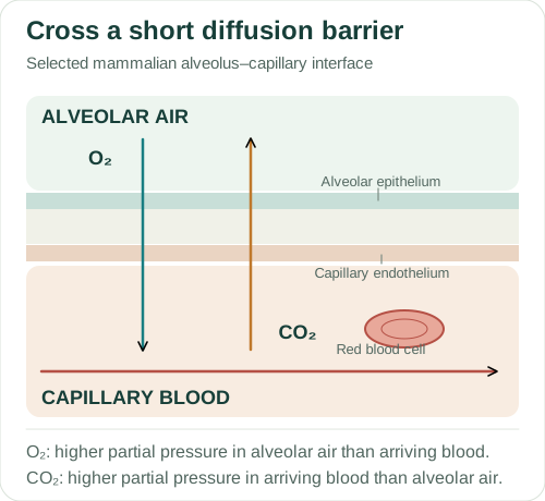
At a mammalian alveolus, oxygen moves net from air to pulmonary capillary blood when alveolar oxygen partial pressure is higher. Carbon dioxide moves net the other way when its partial pressure is higher in arriving blood. Thin alveolar epithelium and capillary endothelium create a short diffusion barrier, and many alveoli provide a large total area. The figure separates the layers but simplifies their intervening material.
Ventilation replaces alveolar gas; blood flow brings blood to the surface and takes it away. Haemoglobin binds oxygen reversibly, enabling much more oxygen to be carried than by dissolution in plasma alone. Loading and unloading depend on oxygen partial pressure and haemoglobin properties. Increased carbon dioxide can reduce oxygen affinity in the Bohr effect, assisting unloading at active tissues.
During quiet inspiration, the diaphragm contracts and flattens and the external intercostal muscles help raise the ribs. Thoracic volume increases, lowering pressure within the lungs below atmospheric pressure, so air enters. Quiet expiration mainly follows relaxation and elastic recoil. Forced expiration recruits additional muscles. Oxygen crossing the barrier is diffusion; air moving in bulk during ventilation is a different process.
About this diagram
Selected interface, not a lung section or complete barrier ultrastructure. Basement membranes and intervening layers are simplified. Arrows describe net gas movement under the stated partial-pressure gradients; haemoglobin loading is explained in the text.
Countercurrent exchange in fish gills
Gill filaments carry many lamellae, providing a large exchange area with a short route between water and blood. In countercurrent exchange, water and blood flow in opposite directions. At positions along a lamella, water can remain at a higher oxygen concentration than the adjacent blood. This maintains a favourable local gradient over much of the exchange surface.
Do not compare the oxygen-rich water entering one end with blood entering the other end as if they meet at the same location. A countercurrent explanation needs paired values at the same position. Opposite flow direction alone is not the whole mechanism; the maintained local difference is what allows oxygen to continue entering the blood. Ventilation across the gills keeps water moving through this arrangement.
Mammalian double circulation
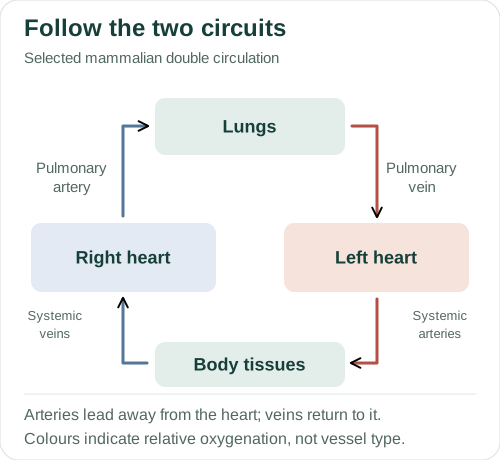
In mammalian double circulation, the right side of the heart sends blood through the pulmonary circuit to the lungs. Blood returns to the left side, which sends it through the systemic circuit to body tissues. It then returns to the right side. Passing through the heart between circuits allows pressure to be raised again for systemic flow while pulmonary pressures remain lower.
An artery carries blood away from the heart; a vein carries it towards the heart. The pulmonary artery therefore carries relatively deoxygenated blood, while pulmonary veins return relatively oxygenated blood. Vessel names are defined by direction, not by oxygen content. In the figure, blue denotes lower oxygenation; human blood is not blue.
Arterial walls contain elastic tissue and muscle suited to pressure and flow control. Capillaries provide short exchange distances; venous return operates at lower pressure and can be assisted by valves and surrounding muscle activity. During the cardiac cycle, valves open or close in response to pressure differences, limiting backflow. Cardiac output is heart rate multiplied by stroke volume; keep the time and volume units consistent.
About this diagram
Flow map, not anatomical chamber positions. Heart chambers, valves and individual vessels are omitted. Red and blue indicate relatively higher and lower oxygenation in these selected circuits; neither colour means blood is literally blue.
Water movement through xylem
Water evaporates from moist mesophyll surfaces and diffuses as vapour through stomata. This lowers water potential in the leaf and draws water from the xylem. Cohesion between water molecules helps transmit tension through the water column; adhesion to vessel walls also contributes. Xylem vessels are largely dead, hollow conduits whose lignified walls resist collapse under tension.
Evaporation drives the transpiration stream. There is no ATP-driven pump inside the dead xylem vessels. Uptake of particular mineral ions by living root cells can involve active transport, but that does not make the entire upward water column an ATP-pumped flow. A potometer estimates water uptake, which is often used as a proxy for transpiration under suitable conditions. It does not directly measure all water lost from leaves.
Humidity, temperature, air movement and stomatal opening affect water loss. Warming a leaf may increase evaporation, but severe water stress can close stomata and restrict water loss. The effect observed therefore depends on the experimental conditions and the plant’s response.
Translocation in phloem
Phloem transports assimilates, especially sucrose, through living sieve-tube elements supported by companion cells. A source supplies assimilate, for example a photosynthesising leaf. A sink uses or stores it, such as a growing root or developing fruit. An organ can change role: a storage organ can become a source when stored material is mobilised.
In the pressure-flow explanation, sucrose loading at a source lowers phloem water potential. Water enters from nearby xylem, raising hydrostatic pressure. Unloading at a sink contributes to a lower pressure, allowing bulk flow along a pressure gradient. Loading in the commonly taught active-loading route depends on metabolism in living cells. Distinguish that energetic dependence from the pressure gradient immediately driving bulk flow.
Translocation can occur in different directions in different sieve tubes because the positions of sources and sinks differ. “Phloem always moves down” therefore fails for a sink above its source. Xylem water flow and phloem assimilate flow have different driving mechanisms.
Worked examples
1. Compare two cubes
Find surface-area-to-volume ratios for cubes of side 1 mm and 2 mm.
- For side 1 mm: area = 6 × 1² = 6 mm²; volume = 1³ = 1 mm³.
- For side 2 mm: area = 6 × 2² = 24 mm²; volume = 2³ = 8 mm³.
- Ratios are 6/1 = 6 mm⁻¹ and 24/8 = 3 mm⁻¹.
Doubling side length halves the ratio, from 6 to 3 mm⁻¹.
The larger cube has more total area but less area per unit volume. State the dimensional ratio as well as any simplified numerical comparison.
2. Calculate cardiac output
Heart rate is 72 beats min⁻¹ and stroke volume is 70 cm³ beat⁻¹. Find cardiac output in dm³ min⁻¹.
- 72 × 70 = 5040 cm³ min⁻¹.
- 1000 cm³ = 1 dm³.
- 5040 ÷ 1000 = 5.04 dm³ min⁻¹.
Cardiac output is 5.04 dm³ min⁻¹ under the supplied conditions.
The beat units cancel. This is volume pumped per time, not a blood velocity or the total volume in the circulation.
3. Explain flow to a growing shoot
A growing shoot above a mature leaf imports sucrose. Explain why an upward phloem flow is consistent with pressure-flow transport.
- The mature leaf acts as a source and the growing shoot as a sink.
- Source loading and water entry can raise phloem pressure.
- Flow follows the source-to-sink pressure difference, not a rule requiring downward movement.
Sucrose can move upwards in phloem from the leaf source to the shoot sink as bulk flow down a pressure gradient.
The position of the organs establishes direction. The mechanism still requires the appropriate source loading, sink unloading and connected transport tissue.
These are original teaching examples, not reproduced exam questions or an official mark scheme.
Common errors
A bigger organism has a larger surface-area-to-volume ratio.
For similar geometry, volume grows faster than area as size increases.
All arteries carry oxygenated blood.
Arteries lead away from the heart; the pulmonary artery is a counterexample to the oxygenation claim.
Ventilation is oxygen diffusion into blood.
Ventilation moves air in bulk; gas exchange crosses the barrier by diffusion.
Phloem can only move downwards.
Its direction depends on the connected source and sink.
Check your understanding
Try the questions before opening the answers.
1. How does blood flow help oxygen enter at an alveolus?
It brings relatively oxygen-poor blood and carries oxygen away after loading, helping maintain the local partial-pressure gradient.
2. What comparison would demonstrate countercurrent exchange?
Compare water and adjacent blood at the same positions along a lamella, showing a favourable oxygen difference maintained as their flows run in opposite directions.
3. Why is potometer uptake not necessarily identical to transpiration?
Some absorbed water can be retained or used by the plant, and apparatus leaks or changing conditions can affect uptake. The interpretation relies on suitable controls and assumptions.
Practice for this topic
- Surface area to volume ratio
- Rates and graph gradients
- Units and standard form
- Apply to a case: Gas exchange and circulation: linking measurements
- Practical guide: Dissection: observations and explanations
Connect this topic to an AQA essay title · Use official papers and mark schemes
Glossary
- Partial pressure
- Contribution of a gas to pressure; differences help describe gas-exchange direction.
- Transpiration
- Loss of water vapour from a plant, mainly through stomata.
- Translocation
- Transport of assimilates through phloem between sources and sinks.
References
References used for the explanations and diagrams, checked on 4 October 2026. The diagrams are schematics and are not to scale. Open “About this diagram” for the assumptions and details left out.